Узбекистан, Андижан

СДЕЛАЙТЕ СВОИ УРОКИ ЕЩЁ ЭФФЕКТИВНЕЕ, А ЖИЗНЬ СВОБОДНЕЕ
Благодаря готовым учебным материалам для работы в классе и дистанционно
Скидки до 50 % на комплекты
только до
Готовые ключевые этапы урока всегда будут у вас под рукой
Организационный момент
Проверка знаний
Объяснение материала
Закрепление изученного
Итоги урока
Был в сети 01.12.2023 15:14
Ашуров Маъруфжон Абдумуталибович
Доцент
45 лет
Местоположение
Специализация
Clinical case of perforation treatment using calcium silicate cement and internal matrix.
Категория:
Право
15.01.2022 17:13

Просмотр содержимого документа
«Clinical case of perforation treatment using calcium silicate cement and internal matrix.»
CLINICAL CASE OF PERFORATION TREATMENT USING CALCIUM SILICATE CEMENT AND INTERNAL MATRIX.
Annotition: Perforations can occur for various reasons, such as caries, resorption, or iatrogenic trauma, such as during access creation, orifice preparation, post space preparation, or during retrieval of broken instruments. Perforations can be detected at the diagnostic stage during an x-ray examination. In the image in the area of perforation, there will be material going beyond the dentin towards the periodontal ligament.
Keywords: Restoration, Clinical case, cement, composite material, formula.
Perforation is the result of a procedural error that can occur in endodontics and affect the prognosis of root canal treatment. Perforation is the second most common cause of failure in endodontics, with Ingl attributing about 9.6% of failure to endodontics to perforation.
Perforations can occur for various reasons, such as caries, resorption, or iatrogenic trauma, such as during access creation, orifice preparation, post space preparation, or during retrieval of broken instruments. Perforations can be detected at the diagnostic stage during an x-ray examination. In the image in the area of perforation, there will be material going beyond the dentin towards the periodontal ligament.
The prognosis of perforation depends on various factors, such as its location, the time elapsed before starting treatment, the ability to close the defect, and microbial contamination. The key factor in determining the degree of complexity of the treatment is the level of perforation occurrence. If the defect is located in the apical third (complication of instrumentation), restoration will be quite difficult. When it is located in the coronal third of the canal (i.e., perforation during access formation) or in the furcation, treatment is considered readily available.
The immediate restoration of perforation is a priority over the delayed one, because the latter can cause destruction of the periodontium, which will lead to endodontic and periodontal lesions, which will complicate treatment. A variety of materials for the treatment of perforations are presented: amalgam, super-ethoxybenzoic acid (EBA), cement, composite material and the most recently used mineral trioxide aggregate (MTA).
MTA was introduced more than 20 years ago as a material for filling root canals. It includes a mixture of 75% Portland cement, 20% bismuth oxide, 5% gypsum and trace amounts of silicone dioxide, calcium oxide, magnesium oxide, potassium sulfate and sodium sulfate. There are 2 types of MTA: gray and white. The gray form includes calcium oxide, dicalcium silicate, tricalcium silicate, tricalcium aluminate, tetracalcium alumoferrite, and calcium sulfate dehydrate, while the white formula lacks tetracalcium aluminoferrite. Currently, MTA is used for various clinical procedures such as apexification, pulp capping, retrograde filling and pulpotomy. MTA has good adhesion to the dentinal surface, which makes the material resistant to shear forces, which is necessary for perforations in the furcation area.
This article describes a case of perforation of the distal-lingual area of the pulp chamber floor of the mandibular permanent molar with its MTA filling with an internal matrix.
Clinical case. A 35-year-old patient presented to the Department of Conservative Dentistry and Endodontics at the Dental Institute, Mathura, Delhi, India, with the main complaint of pain and swelling in the region of the mandibular chewing teeth on the left. This happened after his treatment in a private clinic 2 days ago.
Clinical examination revealed the presence of gingival tissue in the cavity of the left first lower molar. (Image 1).

Image 1. Clinical view before treatment showing perforation of the distal-lingual portion of the pulp chamber floor of the lower first molar on the left.
X-ray examination showed perforation of the distal-lingual area of the bottom of the pulp chamber of the lower first molar on the left. Also, proximal caries was found on the adjacent premolar (Figure 2).
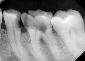
Image 2. Intraoral periapical view showing perforation of the distal-lingual portion of the pulp chamber floor of the lower first molar on the left.
After local anesthesia and placement of a rubber dam, access was created and canals found. Using an apex locator (Root ZX, J Morita, morita.com), the working length was set 1 mm shorter than the radiographic apex and confirmed by radiograph (Image 3).

Image 3. X-ray determination of working length.
The canal was treated with nickel-titanium files (HyFlex ™ CM, Coltene, coltene.com), drug treatment with 5.25% sodium hypochlorite solution and saline solution. The canals were enlarged to 25/04 in the mesial canals (mesiobuccal and mesio-oral) and to 30/04 in the single distal canal. A small portion of synthetic collagen material (CollaTape®, Zimmer Biomet Dental, zimmerbiometdental.com) was gently condensed to cover the furcation area. Gray MTA (MTA Angelus® [Gray], Angelus, angelusdental.com) was prepared according to the manufacturer's instructions and placed on the CollaTape barrier. To avoid the penetration of MTA into the canal, gutta-percha pins were inserted (Image 4).
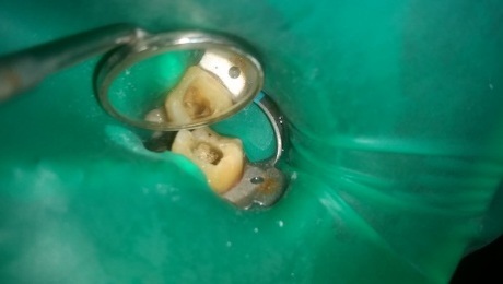
Image 4. Clinical view showing the position of the MTA after matrix insertion.
A damp cotton ball was applied over the MTA to speed up the set of the material, and the access cavity was closed with a temporary filling material (Cavit ™, 3M Oral Care, 3m.com). The location of the MTA was confirmed radiograp hically (Figure 5).

Image 5. X-ray showing the location of the MTA in the perforation of the pulp chamber floor.
The patient returned the next day. The temporary material was removed and the condition of the MTA was assessed. CT scan obtained (Image 6), obturation with single-cone technique was performed using a sealer ((AH Plus®, Dentsply Sirona, dentsplysirona.com) (Image 7).

Image 6. Computed tomography.

Image 7. X-ray image obtained after obturation of the canals with gutta-percha.
The access cavity was filled with composite and the tooth was restored with a metal-ceramic crown (Image 8).

Image 8. X-ray image obtained after fixation of the metal-ceramic crown.
Discussion. This article describes the non-surgical reconstruction of the perforation of the distal-lingual area of the pulp chamber floor of the mandibular molar using MTA biomaterial using an internal matrix. Factors such as the level and location of the lesion, its size and the time elapsed from the appearance of the defect to its recovery affect the prognosis of the treatment of perforation of the furcation. The more time passed before the start of treatment, the worse the prognosis will be.
In the presented case, the perforation of the pulp chamber floor was not extensive and the time between the onset of the defect and the treatment was only 2 days, so the treatment should be successful both clinically and radiologically.
To achieve success in treatment, a dense obturation of the perforated area is important to close the source of contamination and preserve the periodontal structures for optimal healing. A barrier (matrix) technique has been proposed for the treatment of perforation, in which a sterile and non-irritating matrix is made of a material such as hydroxyapatite or calcium sulfate, or resorbable collagen, on top of which an MTA biomaterial is placed.
MTA showed less leakage compared to amalgam and "Super EBA", it is also less cytotoxic and neurotoxic than other restorative materials. Despite the advantages of MTA, it can be inconvenient to use, as it requires closure with a damp cotton ball for 3-4 hours before hardening. Therefore, the endodontic treatment protocol requires termination of the procedure and re-appointment of the patient after the material has hardened, which is often unexpected for both the patient and the practitioner.
Conclusion. The use of an internal matrix of resorbable collagen will provide good control over the compaction of the MTA over the matrix. If in this case the die was placed incorrectly, the sealing would be loose. Consequently, the use of a synthetic matrix of collagen material allowed for the correct placement of the MTA, which in turn increased the tightness. Further study is needed to incorporate the internal matrix fitting approach into a standardized method so that the matrix can be correctly positioned every time it is used.
List of used literature
Anatoly Pavlovich Levitsky, Irina Konstantinovna Mizina. Plaque. K .: Zdorov'ya, 1987. - 80 p. (2nd ed., Revised and enlarged) Series: Library of the Practitioner
Mitchell L. - Fundamentals of Orthodontics. M: GEOTAR-Media, 2017, 376 pages (2nd ed.) Translated from English edited by prof. Yu.M. Malygina ISBN 978-5-9704-4231-9
Peter Dawson. Functional occlusion. From temporomandibular joint to smile planning. M .: Practical medicine, 2016.- 592 p. ISBN 978-5-98811-338-6
R. A. Fadeev, N. V. Zubkova, V. V. Boyko, S. A. Komchenkov, L. L. Komchenkova. To an assistant of an orthodontist dentist: A textbook for dental assistants, students of dental faculties of universities, interns and clinical residents, students of medical schools. SPb .: OOO "MEDI publishing house", 2009. - 208 p. ISBN 978-5-91170-031-7
Vodolatsky M.P. - Orthodontics. Tutorial. Edited by Professor M.P. Vodolatsky Stavropol. SGMA, 2005 .-- 200 p. ISBN 5-89822-060-7


Вебинар для учителей
Свидетельство об участии БЕСПЛАТНО!
Полезное для учителя

Реализация образовательных программ осуществляется с применением исключительно электронного обучения и ДОТ