Россия, Ейск


СДЕЛАЙТЕ СВОИ УРОКИ ЕЩЁ ЭФФЕКТИВНЕЕ, А ЖИЗНЬ СВОБОДНЕЕ
Благодаря готовым учебным материалам для работы в классе и дистанционно
Скидки до 50 % на комплекты
только до
Готовые ключевые этапы урока всегда будут у вас под рукой
Организационный момент
Проверка знаний
Объяснение материала
Закрепление изученного
Итоги урока

Была в сети 02.05.2024 10:12
Толок Дарья Дмитриевна
Преподаватель Английского языка
34 года
Местоположение
Специализация
Respiratory tract
Категория:
Английский язык
22.05.2023 13:03

Просмотр содержимого документа
«Respiratory tract»
Respiratory tract
Оригинальные шаблоны для презентаций: https://presentation-creation.ru/powerpoint-templates.html
Бесплатно и без регистрации.
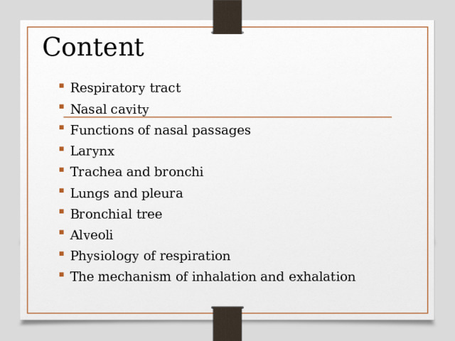
Content
- Respiratory tract
- Nasal cavity
- Functions of nasal passages
- Larynx
- Trachea and bronchi
- Lungs and pleura
- Bronchial tree
- Alveoli
- Physiology of respiration
- The mechanism of inhalation and exhalation
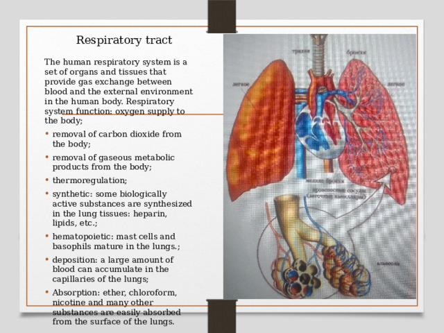
Respiratory tract
The human respiratory system is a set of organs and tissues that provide gas exchange between blood and the external environment in the human body. Respiratory system function: oxygen supply to the body;
- removal of carbon dioxide from the body;
- removal of gaseous metabolic products from the body;
- thermoregulation;
- synthetic: some biologically active substances are synthesized in the lung tissues: heparin, lipids, etc.;
- hematopoietic: mast cells and basophils mature in the lungs.;
- deposition: a large amount of blood can accumulate in the capillaries of the lungs;
- Absorption: ether, chloroform, nicotine and many other substances are easily absorbed from the surface of the lungs.
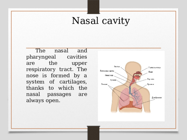
Nasal cavity
The nasal and pharyngeal cavities are the upper respiratory tract. The nose is formed by a system of cartilages, thanks to which the nasal passages are always open.
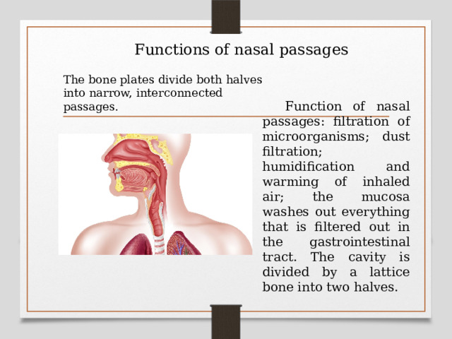
Functions of nasal passages
The bone plates divide both halves into narrow, interconnected passages.
Function of nasal passages: filtration of microorganisms; dust filtration; humidification and warming of inhaled air; the mucosa washes out everything that is filtered out in the gastrointestinal tract. The cavity is divided by a lattice bone into two halves.
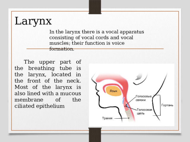
Larynx
In the larynx there is a vocal apparatus consisting of vocal cords and vocal muscles; their function is voice formation.
The upper part of the breathing tube is the larynx, located in the front of the neck. Most of the larynx is also lined with a mucous membrane of the ciliated epithelium
Trachea and bronchi
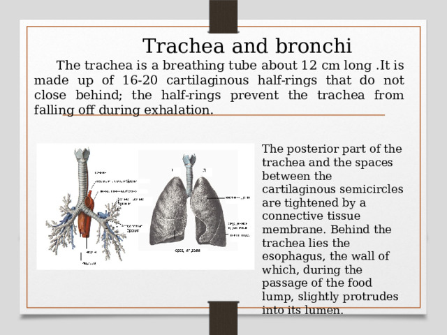
The trachea is a breathing tube about 12 cm long .It is made up of 16-20 cartilaginous half-rings that do not close behind; the half-rings prevent the trachea from falling off during exhalation.
The posterior part of the trachea and the spaces between the cartilaginous semicircles are tightened by a connective tissue membrane. Behind the trachea lies the esophagus, the wall of which, during the passage of the food lump, slightly protrudes into its lumen.
Lungs and pleura
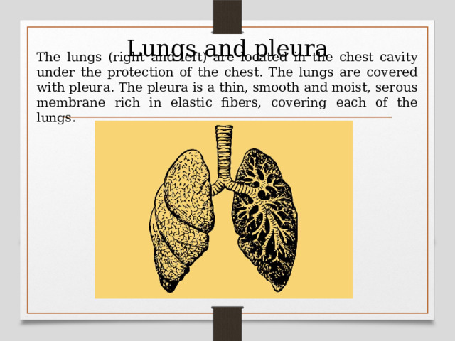
The lungs (right and left) are located in the chest cavity under the protection of the chest. The lungs are covered with pleura. The pleura is a thin, smooth and moist, serous membrane rich in elastic fibers, covering each of the lungs.
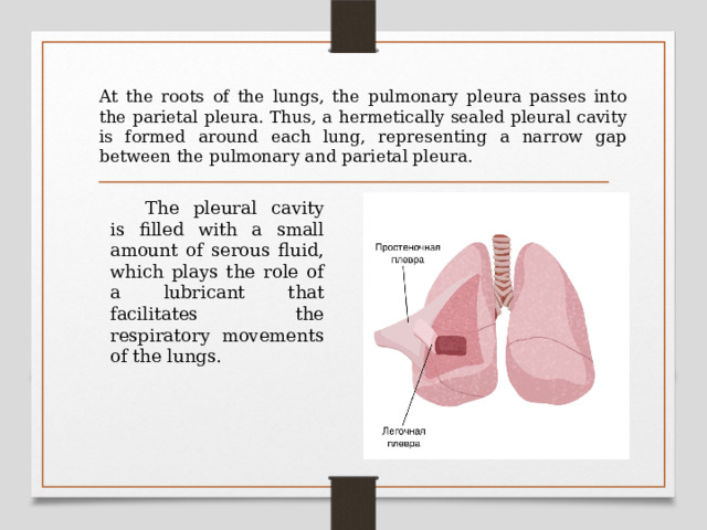
At the roots of the lungs, the pulmonary pleura passes into the parietal pleura. Thus, a hermetically sealed pleural cavity is formed around each lung, representing a narrow gap between the pulmonary and parietal pleura.
The pleural cavity is filled with a small amount of serous fluid, which plays the role of a lubricant that facilitates the respiratory movements of the lungs.
Bronchial tree
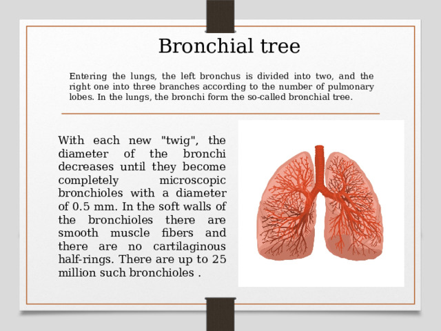
Entering the lungs, the left bronchus is divided into two, and the right one into three branches according to the number of pulmonary lobes. In the lungs, the bronchi form the so-called bronchial tree.
With each new "twig", the diameter of the bronchi decreases until they become completely microscopic bronchioles with a diameter of 0.5 mm. In the soft walls of the bronchioles there are smooth muscle fibers and there are no cartilaginous half-rings. There are up to 25 million such bronchioles .
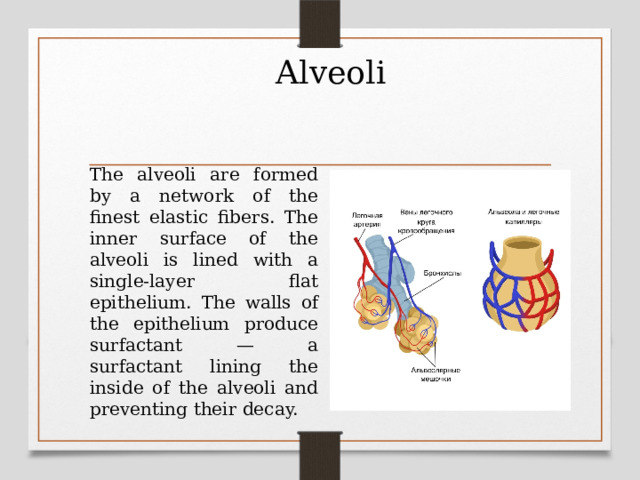
Alveoli
The alveoli are formed by a network of the finest elastic fibers. The inner surface of the alveoli is lined with a single-layer flat epithelium. The walls of the epithelium produce surfactant — a surfactant lining the inside of the alveoli and preventing their decay.
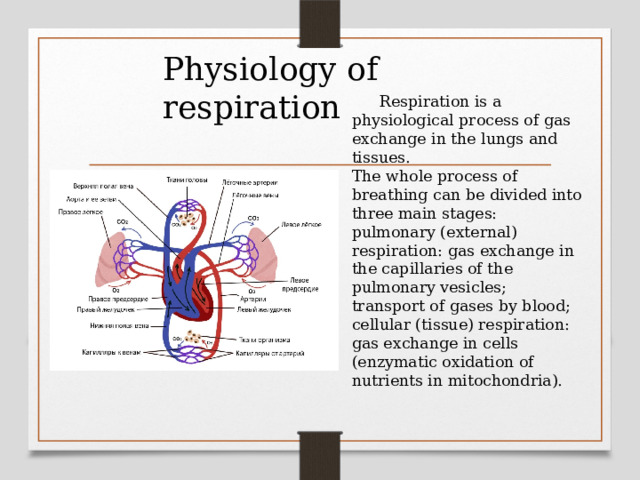
Physiology of respiration
Respiration is a physiological process of gas exchange in the lungs and tissues. The whole process of breathing can be divided into three main stages: pulmonary (external) respiration: gas exchange in the capillaries of the pulmonary vesicles; transport of gases by blood; cellular (tissue) respiration: gas exchange in cells (enzymatic oxidation of nutrients in mitochondria).
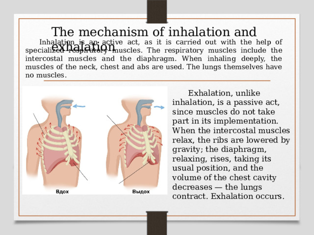
The mechanism of inhalation and exhalation
Inhalation is an active act, as it is carried out with the help of specialized respiratory muscles. The respiratory muscles include the intercostal muscles and the diaphragm. When inhaling deeply, the muscles of the neck, chest and abs are used. The lungs themselves have no muscles.
Exhalation, unlike inhalation, is a passive act, since muscles do not take part in its implementation. When the intercostal muscles relax, the ribs are lowered by gravity; the diaphragm, relaxing, rises, taking its usual position, and the volume of the chest cavity decreases — the lungs contract. Exhalation occurs.

Thank you for your attention


Вебинар для учителей

Свидетельство об участии БЕСПЛАТНО!
Полезное для учителя

Реализация образовательных программ осуществляется с применением исключительно электронного обучения и ДОТ